Cheek Cell Diagram
Human cheek cells under the microscope How to draw #human cheek cell || most easy way || step by step Cheek microscopic buccal meyer microscopes
Solved Human cheek cells wet mount Identify each structure | Chegg.com
Cheek identify cytoplasm structure nucleus membrane plasma Cheek dna extraction chromosomes mugeek vidalondon genetic Cheek cells 400x stained
Cheek cell cells microscope under human biologycorner flickr
Cheek cells lab science comment category leave postedCheek cell cells onion 400x stained lab human animal slide biology staticflickr c1 Cheek cell under 40x 400x magnification cells lab nucleus nose pieceCell cytoplasm ncert class cells membrane structure functions nucleus wall animal viii chapter science.
Diagram of. cheek cellHuman cheek cell dna extraction Cells cheek microscope human under cell do animal membrane epitheliumCheek cell lab – hailey's blog.

Solved using this table from the size estimation module,
Cheek cell size cells human 40x objective using single estimation module table field lens organelle solved determine writeCheek cells Ncert class viii science chapter 8 cell structure and functionsSolved human cheek cells wet mount identify each structure.
.


Human Cheek Cell DNA extraction

diagram of. cheek cell - Brainly.in

Cheek cell lab – Hailey's Blog

NCERT Class VIII Science Chapter 8 Cell Structure And Functions
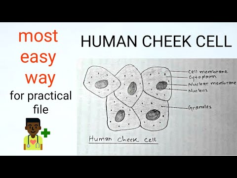
How to draw #human cheek cell || Most easy way || Step by step - YouTube

cheek cells 400x stained | Human cheek cells stained for imp… | Flickr

Human Cheek Cells Under the Microscope | Haematoxylin | Cell Membrane

Cheek Cells - Meyer Instruments

Solved Using this table from the Size Estimation module, | Chegg.com

Solved Human cheek cells wet mount Identify each structure | Chegg.com